Tag Archives: Hepatitis C virus

Baruch Blumberg: The Hepatitis B Virus and Vaccine

Hepatitis B virus (HBV), one of mankind’s most important pathogens, infects about 2 billion people worldwide, and more than 500 million individuals are life-long carriers of the virus; with most in Asia. HBV causes acute and chronic cirrhosis, as well as hepatocellular carcinoma. In point of fact, HBV is the 10th leading cause of death in the world! The serendipitous discovery of HBV, and the development of the first HBV vaccine, happened as follows. [See Note 1 for a brief review of the remarkable HBV replication strategy].

In the early 1940’s, during World War II, British doctor, F. O. MacCallum, was the first to suggest that an infectious agent might cause hepatitis. MacCallum was assigned to produce a yellow fever vaccine for British soldiers. That was how he happened to notice that soldiers tended to come down with hepatitis a few months after receiving the yellow fever vaccine.

It was fortunate that MacCallum also knew of hepatitis cases in children who received inoculations of serum from patients convalescing from measles and mumps (a means of protection against those viruses before vaccines were available), and of hepatitis cases in blood transfusion recipients, and of cases following treatments with unsterilized reused syringes.

To explain these coincidences, MacCallum hypothesized that hepatitis might be transmitted by a factor in human blood. And, since hepatitis could be transmitted by inoculation with serum that had been filtered, MacCallum proposed that the hepatitis factor might be a virus. [In 1947 MacCallum reported that hepatitis could be spread by food and water that had been contaminated with fecal material, as well as by blood. He coined the term hepatitis A for the form of the disease spread by food and water, and hepatitis B for the form transmitted via blood.] See Aside 1.

[Aside 1: The following episode, described in MacCallum’s own words (1), occurred in England during World War II: “One day in 1942, I received a message to go to Whitehall to see one of the senior medical advisers and when I arrived I was asked ‘What is this yellow fever vaccine and how dangerous is it?’ After explaining its constitution and the possibility of a mild reaction four to five days after inoculation, I was told that the Cabinet was at that moment debating whether or not Mr. Churchill should be allowed to go to Moscow, which he wished to do in a few days’ time. The yellow fever vaccine was theoretically essential before he could fly through the Middle East, but I explained that no antibody would be produced before seven to ten days so that there would be little point in giving the vaccine. It was finally decided that the vaccine would not be used, and the administrators would take care of the situation. Several months later I received an irate call from the Director of Medical Services of the RAF, who had been inoculated with the same batch of vaccine which would have been used for Mr. Churchill, and was informed that the D. G. had spent a very mouldy Christmas with hepatitis about 66 days after his inoculation…I will leave it to you to speculate on what might possibly have been the effect on the liver of our most famous statesman and our ultimate fate if  he had received the icterogenic vaccine.”]

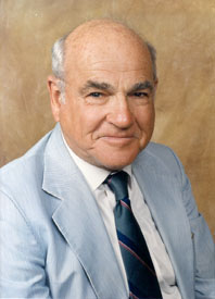
With the advent of cell culture in the 1950s, researchers hoped that a hepatitis agent might soon be cultivated in vitro. Nonetheless, HBV was not discovered until 1966. What’s more, the discovery did not involve growing the virus in cell culture. And, reminiscent of the case of MacCallum above, the discovery was made by a researcher, Baruch S. Blumberg, who was not even working on hepatitis. Rather, Blumberg was interested in why individuals varied in their susceptibilities to various illnesses.

Nobel Laureate Baruch Blumberg

Blumberg sought to answer that question by identifying possibly relevant genetic differences between population groups, which, in the pre-molecular biology era, might be revealed by differences in their blood proteins. Thus, in the early 1950s, Blumberg, then working at the NIH, began collecting a panel of blood samples from diverse populations throughout the world.

Blumberg looked for serum protein variations (i.e., serum protein polymorphisms) by asking if sera from multiply-transfused individuals (defined by Blumberg as persons who received 25 units of blood or more) might contain antibodies that reacted with proteins in the serum samples of his panel. His rational, in his own words, was as follows: “We decided to test the hypothesis that patients who received large numbers of transfusions might develop antibodies against one or more of the polymorphic serum proteins (either known or unknown) which they themselves had not inherited, but which the blood donors had (2).” In other words, patients who received multiple transfusions were more likely than others to have antibodies against polymorphic serum proteins in donor blood, and those antibodies might also react with polymorphic serum proteins in the samples from his panel. See Aside 2.

[Aside 2: Blumberg used the Ouchterlony double-diffusion agar gel technique in these experiments. Serum samples to be tested against each other were placed in opposite wells of a gel. The proteins they contained could then diffuse through the gel. Antigen-antibody complexes that formed between the two samples appeared as white lines in the gel.]

Hemophilia and leukemia patients were well-represented in Blumberg’s collection of serum samples from multiply-transfused individuals. And, a serendipitous aspect of Blumberg’s experimental approach was that he used these samples to probe for serum protein polymorphisms in samples from geographically diverse populations. Thus it happened that Blumberg detected a cross-reaction between a New York hemophilia patient’s serum and a serum sample from an Australian aborigine. But what could these two individuals have had in common that might have triggered the cross-reaction?

His curiosity thus aroused, Blumberg and collaborator, Harvey Alter, of the NIH Blood Bank, tested the hemophilia patient’s serum against thousands of other serum samples. Blumberg and Alter may have been surprised to find that whatever the antigen in the Aborigine’s serum was that reacted with the hemophilia patient’s serum, reactivity against that antigen was common (one in ten) in leukemia patients, but rare (one in 1,000) in normal individuals. In any case, because the antigen was first identified in an Australian aborigine, it was termed the Australia antigen.

Bear in mind that Blumberg’s original purpose was to explain why individuals differed in their susceptibilities to various illnesses. Thus, Blumberg at first believed that he detected an inherited blood-protein that predisposes people to leukemia. However, additional experiments showed that the antigen was more common in older individuals than in younger ones; a finding more consistent with the possibility that the antigen might be associated with an infectious agent.

Blumberg’s first clue that the Australia antigen might be associated with hepatitis came to light when he tested serum samples from a 12-year old boy with Down syndrome. The first time that the boy was tested for the Australia antigen, he was negative. However, several months later, when retested, the boy was positive. Moreover, sometime during that interim, the boy also developed hepatitis.

Blumberg, and other researchers, carried out additional experiments, which confirmed that the Australia antigen indeed associated with hepatitis. In addition, the antigen was more frequently detected in hepatitis sufferers than in individuals with other liver diseases. Thus, the Australia antigen was a marker of hepatitis in particular and not of liver pathology in general. See Aside 3.

[Aside 3: Blumberg had a personal reason motivating him to identify the cause of hepatitis. His technician (later Dr. Barbara Werner) became ill with hepatitis, which she almost certainly acquired in the laboratory. Fortunately, she underwent a complete recovery.]

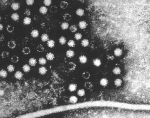
In 1970, British pathologist David Dane and colleagues at Middlesex Hospital in London, and K. E. Anderson and colleagues in New York, provided corroborating evidence  that hepatitis is an infectious disease. Using electron microscopy, they observed 42-nm “virus-like particles” in the sera of patients who were positive for the Australia antigen. In addition, they saw these same particles in liver cells of patients with hepatitis.

What then is the Australia antigen? Actually, it is the surface protein of the 42-nm HBV particles; now known as the hepatitis B surface antigen (HBsAg). Since HBV particles per se were described for the first time by David Dane, they are sometimes referred to as Dane particles.

Now we can explain Blumberg’s early finding, that individuals who received multiple transfusions (e.g., leukemia and hemophilia patients) were more likely than the general population to have antibodies against the Australia antigen. Those individuals were more likely than the general population to have received donated blood and, thus, were more likely to have been recipients of blood contaminated with HBV. At that time, a large percentage of the blood supply came from paid donors, at least some of whom were syringe-sharing, intravenous drug abusers and, consequently, more likely than most to be HBV carriers. In 1972 it became law in the United States that all donated blood be screened for HBV. See Note 2.

But it was important to protect all people from HBV; not just transfusion recipients. In 1968, Blumberg, now at the Fox Chase Cancer Center in Philadelphia, and collaborator Irving Millman, hypothesized that HBsAg might provoke an immune response that would protect people against HBV and, consequently, that a vaccine could be made using HBsAg purified from the blood of HBV carriers. In Blumberg’s own words: “Irving Millman and I applied separation techniques for isolating and purifying the surface antigen and proposed using this material as a vaccine. To our knowledge, this was a unique approach to the production of a vaccine; that is, obtaining the immunizing antigen directly from the blood of human carriers of the virus (1).”  The Fox Chase Cancer Center filed a patent for the process in 1969.

Blumberg was willing to share his method and the patent with any pharmaceutical company willing to develop an HBV vaccine for widespread use. Nonetheless, the scientific establishment was somewhat slow to accept his experimental findings and his proposal for making the vaccine. Then, in 1971, Merck accepted a license from Fox Chase to develop the vaccine. In 1982, after more years of research and testing, Maurice Hillman (3) and colleagues at Merck turned out the first commercial HBV vaccine (“Heptavax”). Producing an HBV vaccine, without having to cultivate the virus in vitro, was considered one of the major medical achievements of the day. See Notes 3 and 4.

The consequences of Blumberg’s vaccine were immediate and striking. For instance, in China the rate of chronic HBV infection among children fell from 15% to around 1% in less than a decade. And, in the United States, and in many other countries, post-transfusion hepatitis B was nearly eradicated.

Moreover, Blumberg’s HBV vaccine was, in a real sense, the world’s first anti-cancer vaccine since it prevented HBV-induced hepatocellular carcinoma, which accounts for 80% of all liver cancer; the 9th leading cause of death. Jonathan Chernoff (the scientific director of the Fox Chase Cancer Center, where Blumberg spent most of his professional life) stated: “I think it’s fair to say that Barry (Blumberg) prevented more cancer deaths than any person who’s ever lived (4).”

In 1976 Blumberg was awarded the Nobel Prize in Physiology or Medicine for “discoveries concerning new mechanisms for the origin and dissemination of infectious diseases.” He shared the award with Carlton Gajdusek, who won his portion for discoveries regarding the epidemiology of kuru (5). See Note 5.

Blumberg claimed that saving lives was the whole point of his career. “This is what drew me to medicine. There is, in Jewish thought, this idea that if you save a single life, you save the whole world, and that affected me (7).” See Aside 4.

[Aside 4: Blumberg received his elementary school education at an orthodox yeshiva in Brooklyn, and he attended weekly Talmud discussion classes until his death. Interestingly, Blumberg graduated from Far Rockaway High School in Queens, N.Y.; also the alma mater of fellow Nobel laureates, physicists Burton Richter and Richard Feynman.]

As we’ve seen, Blumberg’s landmark discovery of HBV sprang from a basic study of human genetic polymorphisms. In Blumberg’s own words, “… it is clear that I could not have planned the investigation at its beginning to find the cause of hepatitis B. This experience does not encourage an approach to basic research which is based exclusively on specific-goal-directed programs for the solution of biological problems (1).”

Saul Krugman (Note 4) had this to say about Blumberg’s discovery: “It is well known that Blumberg’s study that led to the discovery of Australia antigen was not designed to discover the causative agent of type B hepatitis. If he had included this objective in his grant application, the study section would have considered him either naïve or out of his mind. Yet the chance inclusion of one serum specimen from an Australian aborigine in a panel of 24 sera that was used in his study of polymorphisms in serum proteins…led to detection of an antigen that subsequently proved to be the hepatitis B surface antigen (1).” See Note 6.

In 1999, Blumberg’s scientific career took a rather curious turn when he accepted an appointment by NASA administrator, Dan Goldin, to head the NASA Astrobiology Institute. There, Blumberg helped to establish NASA’s search for extraterrestrial life. Blumberg also served on the board of the SETI Institute in Mountain View, Calif.

Blumberg passed away on April 5, 2011, at 85 years of age.

Notes:

[Note 1:  HBV is the prototype virus for the hepadnavirus family, which displays the most remarkable, and perhaps bizarre, viral replication strategy known. In brief, in the cell nucleus, the cellular RNA polymerase II enzyme transcribes the hepadnavirus circular, double-stranded DNA genome, thereby generating several distinct species of viral RNA transcripts, all of which are exported to the cytoplasm. The largest of these viral transcripts is the pregenomic RNA; a transcript of the entire circular viral DNA genome, as well as an additional terminal redundant sequence. Remarkably, the pregenomic RNA is then packaged in nascent virus capsids, within which it is reverse transcribed by a virus-encoded reverse transcriptase activity, thereby becoming an encapsulated progeny hepadnavirus double-stranded DNA genome. Thus, reverse transcription is a crucial step in the replication cycle of the hepadnaviruses, as it is in the case of the retroviruses. But, while the retroviruses replicate their RNA genomes via a DNA intermediate, the hepadnaviruses replicate their DNA genomes via an RNA intermediate.]

[Note 2: The highly sensitive radioimmunoassay (RIA) technique, developed by Rosalyn Yallow and Solomon Berson, is the basis for the test that screens the blood supply for the Australia antigen. The story behind this assay is worthy of note here because it is yet another example of serendipity in the progress of science. In brief, Yallow and Berson sought to develop an assay to measure insulin levels in diabetics. Towards that end, they happened to find that radioactively-labeled insulin disappeared more slowly from the blood samples of people previously given an injection of insulin than from the blood samples of untreated patients. That observation led them to conclude that the treated patients had earlier generated an insulin-binding antibody. And, from that premise they hit upon the RIA procedure. Using their insulin test as an example, they would add increasing amounts of an unlabelled insulin sample to a known amount of antibody bound to radioactively labeled insulin. They would then measure the amount label displaced from the antibody, from which they could calculate the amount of unlabelled insulin in the test sample. Their procedure has since been applied to hundreds of other substances. RIA is simpler to carry out and also about 1,000-fold more sensitive than the double-diffusion agar gel procedure that Blumberg used to identify the Australia antigen. Yallow and Berson refused to patent their RIA procedure, despite its huge commercial value. Yallow received a share of the 1977 Nobel Prize in Physiology or Medicine for her role in its development. Berson, died in 1972 and did not share in the award.]

[Note 3: Making Heptavax directly from the blood of human HBV carriers was somewhat hindered because it required a continuing and uncertain supply of suitable donor blood. Moreover, there was concern that even after purifying the HBsAg, and treating it with formalin to inactivate any infectivity, the vaccine might yet contain other live dangerous viruses. Concern increased in the early 1980s with the emergence of HIV/AIDS, since much of the HBV-infected serum came from donors who later developed AIDS. Thus, in 1990 Heptavax was replaced in the United States by a safer genetically engineered (i.e., DNA recombinant) HBV vaccine, which contained no virus whatsoever. That vaccine was the first to be made using recombinant DNA technology. Moreover, it was yet another instance in which Hilleman played a key role in the development of a vaccine (3).]

[Note 4: In 1971, Saul Krugman, working at NYU, was actually the first researcher to make a “vaccine” against HBV. Krugman’s accomplishment began as a straightforward inquiry into whether heat (boiling) might kill HAV (see Note 5). Finding that it did, Krugman repeated his experiments; this time to determine whether boiling might likewise kill HBV in the serum of HBV carriers. As Krugman expected, boiling indeed destroyed HBV infectivity. But, to his surprise, while the heated serum was no longer infectious, it did induce incomplete, but statistically significant protection against challenge with live HBV. Krugman considers his “vaccine” discovery, like Blumberg’s discovery of HBV, to have resulted from “pure serendipity” (1).

Krugman could not answer whether HBsAg per se in his crude vaccine induced immunity. However, Hilleman, in 1975, using purified HBsAg, as per Blumberg’s concept, showed that HBsAg indeed induced immunity against an intravenous challenge with HBV.

Krugman also carried out key studies on the epidemiology of hepatitis, demonstrating that “infectious” (type A) hepatitis is transmitted by the fecal-oral route, while the more serious “serum” (type B) hepatitis is transmitted by blood and sexual contact.

Krugman reputation was somewhat tarnished because he used institutionalized disabled children as test subjects in the experiments that led to his vaccine. While that practice astonishes us today, it was not unheard-of in the day. In any event, it did not prevent Krugman’s election in 1972 as president of the American Pediatric Society, or his 1983 Lasker Public Service Award.]

[Note 5: Gajdusek’s reputation was later sullied when he was convicted of child molestation (5).]

[Note 6: In 1973 and 1974, research groups led by Stephen Feinstone and Maurice Hilleman (3) discovered hepatitis A virus (HAV), a picornavirus.

After the discoveries of HAV and HBV, it became clear that blood samples cleared of HAV and HBV could still transmit hepatitis. In 1983 Mikhail Balayan identified a virus, now known a hepatitis E virus (the prototype of a new family of RNA viruses), as the cause of a non-A, non-B infectious hepatitis (6).

In 1989, a mysterious non-A, non-B hepatitis agent, now known as hepatitis C virus (a flavivirus), was identified by a team of molecular biologists using the cutting-edge molecular biology techniques of the day (8).]

References:

  1. Krugman, S. 1976. Viral Hepatitis: Overview and Historical Perspectives. The Yale Journal of Biology and Medicine 49:199-203.
  1. Blumberg, B, Australia Antigen and the Biology of Hepatitis B, Nobel Lecture, December 13, 1976.
  1. Maurice Hilleman: Unsung Giant of Vaccinology, Posted on the blog April 24, 20143.
  1. Emma Brown (6 April 2011). “Nobelist Baruch Blumberg, who discovered hepatitis B, dies at 85”. The Washington Post.
  1. Carlton Gajdusek, Kuru, and Cannibalism, Posted on the blog April 6, 2015.
  1. Mikhail Balayan and the Bizarre Discovery of Hepatitis E Virus, Posted on the blog May 3, 2016.
  1. Segelken, H. Roger (6 April 2011). “Baruch Blumberg, Who Discovered and Tackled Hepatitis B, Dies at 85”. New York Times.
  1. Choo, Q. L., G. Kuo, A.J. Weiner, L.R. Overby, D.W. Bradley, and M. Houghton. 1989. Isolation of a cDNA clone derived from non-A, non-B viral hepatitis genome. Science 244:359-362.


Mikhail Balayan and the Bizarre Discovery of Hepatitis E Virus

There have been several instances in which medical researchers, for the sake of mankind, allowed themselves to be infected with a potentially deadly pathogen. A well known example involved the discovery that the Aedes aegypti mosquito is the vector for yellow fever (1). Here we consider a less known and slightly bizarre example in which Mikhail S. Balayan, of the Russian Academy of Medical Sciences in Moscow, discovered the hepatitis E virus.

But first, hepatitis refers to an inflammatory disease involving the liver. Four unrelated viruses, hepatitis A, hepatitis B, hepatitis C, and hepatitis E viruses cause epidemic viral hepatitis (see Aside 1). Hepatitis E was initially identified in 1980 as a non-A, non-B infectious hepatitis. The differences between hepatitis A, B, and E virus infections are as follows. Hepatitis A and hepatitis E are similar, insofar as the etiologic agent of each usually gives rise to an acute (i.e., self-limiting) infection and illness. In contrast, hepatitis B and hepatitis C viruses usually give rise to persistent infections that may lead to chronic hepatitis, cirrhosis, and liver cancer. The mortality rate for hepatitis E is generally “only” about 1% to 2%. Yet, hepatitis E is unusual among hepatitis viruses for its severity in pregnant woman, in whom the fatality rate may reach 20%.

[Aside 1: For aficionados, hepatitis A is a picornavirus, hepatitis B is a hepadnavirus (a DNA retrovirus), and hepatitis C is a flavivirus. Hepatitis E-like viruses were originally classified as calciviruses. However, sequencing of their RNA genomes revealed that they are more similar to rubella virus, a togavirus, than to the calciviruses. Yet they are different enough from togaviruses to merit their own family. The prototype is the hepatitis E virus, discovered by Balayan. Like hepatitis A virus, it is spread by the fecal-oral route. Hepatitis E virus is found worldwide, but it is most problematic in developing countries.]

Here then is Balayan’s tale. In 1983 Balayan was investigating an outbreak of non-A, non-B hepatitis in Tashkent; now the capital city of Uzbekistan. Balayan wanted to bring patient samples back to Moscow to study. However, he had no means for refrigerating the samples. Moreover, he may not have had permission from his supervisors to return with the samples. So, he solved his dilemma by a rather extreme form of self sacrifice—he drank a pooled filtrate of patient stool samples. He is said to have made his private inoculum more palatable by first mixing it with yogurt.

Belayan’s efforts were not for naught since, after returning to Moscow, he indeed came down with hepatitis, as he presumably desired. In fact, he became seriously ill. He then began to collect his own stool samples, in which he detected, by electron microscopy, 32 nm virus particles that produced a hepatitis-like illness when inoculated into monkeys. Balayan then observed a virus in the stool of these monkeys that appeared to be identical to the virus in the original patient samples, which he transported in, and recovered from himself.

Hepatitis E Virus
Hepatitis E Virus

Belayan’s virus looked like hepatitis A virus in electron micrographs. But, he could show that it was not hepatitis A virus. He already had antibodies against the hepatitis A virus, and these did not react with the new virus.

Balayan mentions himself in his original report (2), as follows: “Hepatitis E virus (HEV) was first identified in the excreta of an experimentally infected human volunteer and further confirmed by similar findings in clinical specimens from patients with acute jaundice disease different from hepatitis A and B.”

References:

1. The Struggle Against Yellow fever: Featuring Walter Reed and Max Theiler, Posted on the blog May 13, 2014.

2. Balayan, M.S., 1983. Hepatitis E virus infection in Europe: Regional situation regarding laboratory diagnosis and epidemiology. Clinical and Diagnostic Virology 1:1-9.


Zika Virus: Background, Politics, and Prospects

Ebola, MERS, and Hepatitis C viruses dominated virology news during the past year (2015). Now, early in 2016, Zika virus has taken center stage. The reasons are clear. This once seemingly innocuous virus, initially restricted to Equatorial Africa, has of late spread to the Western Hemisphere, and is now suspected (but not proven) to cause microcephaly—an otherwise rare condition in which babies have unusually small heads and incomplete brain development—in transplacentally infected fetuses of infected pregnant woman. Moreover there is evidence which links Zika virus to Guillain–Barré syndrome—a potentially severe autoimmune attack on peripheral nerves that may occur after signs of a viral infection. We begin with some background.

Zika virus is a member of the flavivirus family of plus-strand RNA viruses. The family also includes several notable human pathogens, including yellow fever, dengue, hepatitis C, and West Nile viruses. Like most other flaviviruses, Zika virus too is spread by an arthropod vector; in this instance Aedes mosquitoes. 80% of Zika virus infections are asymptomatic and, prior to recent developments, symptomatic infections were seen as mild, acute febrile illnesses, similar to dengue.

Zika virus was discovered by accident in the Zika Forest of Uganda in 1947. The discovery was made by scientists who had been studying yellow fever. They isolated Zika virus from one of their rhesus macaques, which was suffering from an unknown fever. The following year the same virus was found in Aedes mosquitos from the same Ugandan forest, thus identifying the mosquito as a vector for Zika virus. Zika virus was detected for the first time in humans in 1954, in Nigeria.

The Aedes aegypti mosquito, the Zika virus vector

Until recently, Zika virus infections were rare and were reported only within equatorial Africa and Southeast Asia. Then, in 2007, an outbreak occurred in Yap Island, Micronesia. The Yap Island Zika outbreak was the first one outside of Africa and Asia. None of the Yap Island cases, which included 49 in which Zinka virus was confirmed by the presence of Zinka RNA, resulted in either hospitalization or death.

The Yap Island outbreak was followed by epidemics in Polynesia, Easter Island, the Cook Islands and New Caledonia. The Polynesian outbreak was notable for being the first in which Zika infection was associated with Guillain–Barré syndrome.

Concern over Zika virus was heightened, particularly in the Americas, when, in April 2015, a large and still ongoing outbreak of Zika virus occurred in Brazil. The Brazilian outbreak marked the first appearance of Zika virus in the Western Hemisphere. It is not clear how Zika virus made its way to Brazil, but it is widely believed that the virus made the leap from Polynesia to Brazil during the 2014 World Cup soccer tournament.

Apprehension over Zika virus increased in November 2015 when the virus was isolated from a Brazilian newborn with microcephaly. By December 2015 many more cases of this generally rare disorder were reported. The European Center for Disease Prevention and Control then warned of a possible association between Zika virus infection and congenital microcephaly, and with Guillain–Barré syndrome as well.

More than a million Brazilian people since been infected with Zika virus, and the number of Brazilian children born with microcephaly jumped from 147 in 2014 to nearly 4,000 in 2015. There is no anti-Zika vaccine, nor is there an effective therapy.

The first Zika virus-associated case of microcephaly in the United States occurred in early January 2016 in a baby born in Oahu, Hawaii. The baby and its mother each tested positive for a past Zinka infection; probably acquired in May 2015 when the mother, then pregnant, had been traveling in Brazil.

On January 24, 2016 the World Health Organization warned that Zika virus will likely spread to every nation in the Western Hemisphere (possibly excepting Canada and Chile), since its Aedes aegypti vector can thrive in tropical and sub-tropical climates here. The Aedes mosquito has long been present in the United States, ranging as far north as New York and west into Indiana and Illinois. [An earlier posting reported that Aedes aegypti may have been brought from Africa to the New World by slave ships in 1596 (1). Mosquito larvae, present in the water casks of the sailing ships of the day, also carried yellow fever to the New World.]

Global concern over the Brazilian Zika outbreak was heightened by the fact that Brazil is scheduled to host the Olympic Games this summer, and about 500,000 people are expected to attend from all over the world, including 200,000 Americans. Some of these attendees will, of course, be bringing the virus back to their home countries.

Brazilian officials no doubt are concerned that their Zika outbreak will affect attendance at the upcoming Olympic Games. Consequently, commercial considerations may be one of the motives behind Brazil’s extensive campaign to eradicate its mosquitoes. Unfortunately, standard approaches, such as using insecticides and removing standing water where mosquitoes breed, have not done the job. Thus, the Brazilian Zika outbreak may not be under control by the start of the Olympic Games. [Brazil also experienced more than 1.6 million cases of dengue during 2015, with 863 people dying from the disease, underscoring that the Aedes mosquito vector is not well contained in that country.]

The failure of Brazilian vector-control approaches suggests that new strategies may be needed to contain the outbreak. Apropos that, this past January Colombia began releasing mosquitoes treated with bacteria, which are hoped might limit the mosquitoes’ capacity to spread disease. Note that insecticides have limited effectiveness. Not only are they toxic to humans, but after decades of overexposure to them, many mosquitoes are now resistant.

Zika virus is now present in the continental United States. Thus, it is timely to consider how grave a threat Zika virus might impose here. To that point, consider that yellow fever, dengue and chikungunya viruses are dangerous pathogens that also are spread by Aedes mosquitoes. Yet these viruses are not regarded as important threats in the United States. That is so because our vector control measures have thus far been able to contain them. Those measures might likewise be expected to contain local transmission of Zika virus here.

But, what if Zika virus has a mode of transmission other than via its mosquito vector? To that point, there is a single reported case of Zika transmission via a blood transfusion. Also, it was suggested that Zika virus might have a sexual route of transmission, as per the finding of high levels of the virus in the semen of a man from French Polynesia. In addition, there is a report of an American scientist, Brian D. Foy, who contracted Zika virus while working in Senegal in 2008, and who transmitted the virus to his wife after returning home (2). Serologic analyses of the couple’s convalescent serum confirmed that they had been infected with Zika. Sexual transmission is implicated in this instance since neither Foy nor his wife passed the infection to their children or to other close relatives. Moreover, Foy and his wife observed signs of hematospermia (red–brown fluid in his ejaculate).

Foy notes in his scientific report (2), “If sexual transmission could be verified in subsequent studies, this would have major implications toward the epidemiology of Zika virus and possibly other arthropod-borne flaviviruses.” [Human sexual transmission of an arthropod-borne virus has not yet been documented.] Foy has been trying to get funds to investigate sexual transmission of Zika. However, according to a January 26, 2016 article in the N.Y. Times, the CDC says that the “theoretical risk” of sexual transmission in the above instances is insufficient to justify a warning (and funding?). But, see the following paragraph.

As I’m sitting at my computer on the evening of February 2, 2016, NPR, CNN, BBC News, the N.Y. Times, etc., are reporting a case of Zika virus infection in Texas that appears to have been sexually transmitted. According to the Dallas County Health and Human Services Department, a patient with the Zika virus was infected after having sex with someone who returned from Venezuela, where Zika is circulating. The CDC appears to give credence to the Texas report, since it quickly responded to it by advising men having sex after traveling to these areas to “consider” wearing condoms, and advised pregnant women to avoid “contact with semen” from men recently exposed to the virus.

Sexual transmission will probably account for only a very small fraction of Zika cases, but that isn’t known for certain. As in instances of mosquito-borne transmission, its contribution will depend in part on how long the virus might persist in infected individuals.

Since the vast majority of Zika virus infections are likely transmitted via its mosquito vector, and since Zika virus mainly threatens fetuses infected in utero, the most severe consequences of Zika virus infection can be largely avoided if pregnant women, or women planning to become pregnant, avoid traveling to places where Zika virus remains prevalent (a fact which doesn’t help individuals living in those regions). For that reason, on January 15, 2016, the United States Centers for Disease Control and Prevention (CDC) released a list of countries—Brazil, Colombia, El Salvador, French Guiana, Guatemala, Haiti, Honduras, Martinique, Mexico, Panama, Paraguay, Suriname, Venezuela, and Puerto Rico—where mosquitoes are spreading the Zika virus, and which pregnant women should avoid at this time. On February 1, 2016, the World Health Organization added Costa Rica and Jamaica.

Political and commercial considerations may have been behind the Brazilian minister of tourism taking exception to the CDC’s warning, claiming that measures adapted by Brazilian health authorities are bringing the Zika outbreak under control, and that Brazil is, in fact, a safe destination for pregnant women. The Brazilian health minister added, “Zika virus doesn’t worry us…,” calling it a “benign disease.” Those pronouncements were made despite the fact that Brazilian health authorities were at the same time investigating more than 3,500 cases of microcephaly. But at least some Brazilian health professionals did endorse the CDC announcement.

On February 1, 2016 the World Health Organization took the further step of declaring that Zika virus and its suspected link to birth defects constitute an international public health emergency. Yet the WHO stopped short of advising pregnant women not to travel to affected regions. Some public health experts claimed that the WHO’s silence on that point was more about politics than public health. Any travel ban—even one aimed only at pregnant women—would be embarrassing and costly to Brazil, which is moving ahead with its plans to host the Olympic Games this summer. And, while there have been calls to cancel, postpone, or move the Rio games, the International Olympic Committee (IOC) hasn’t expressed any concerns over the Games taking place as planned.

Meanwhile, the governments of Columbia, El Salvador, Ecuador, and Jamaica have taken the rather extraordinary step of recommending that women avoid getting pregnant until the Zika outbreak might be brought under control in their countries. This advisory was not well received by many El Salvadoran women, especially in view of the strict abortion laws and high levels of sexual violence against women in that country.

And, as I’m putting the final touches on this piece, an article in today’s (February 4, 2016) N.Y. Times reports that the Zika virus/microcephaly link is causing a fierce debate in Brazil over its strict abortion laws; under which abortion is illegal under most circumstances. [Remarkably, Brazil’s strict abortion laws are actually less restrictive than those in other Latin American countries.] Some Brazilian doctors are already seeing pregnant women who are seeking abortions because they fear microcephaly. Yet conservative Brazilian lawmakers actually want to make the restrictions against abortion more stringent than they already are. [The Times article says that their position reflects “the influence of Roman Catholic leaders and the increasingly powerful preachers at the helm of a growing evangelical Christian movement.”] Regardless, individuals on both sides of the debate might be troubled by the fact that microcephaly can not be detected by ultrasound scans until the end of the second trimester, when the “child” is already very much formed. Moreover, the criteria for diagnosing microcephaly are rather non-specific, and it is difficult to predict what its consequences might be.

A crucially important question regarding Zika virus concerns determining its true pathologic potential, particularly its role in microcephaly—a role that is strongly inferred (but not proven) by the geographic and temporal relationship between microcephaly and Zika infection. To that point, no increase in microcephaly has been linked to Zika virus outside of Brazil. For instance, Colombia is the second-most Zika-affected country, with around 20,000 confirmed cases. More than 2,000 of the Columbian cases have been pregnant women. Yet none of their fetuses have been diagnosed with microcephaly.

Did Zika virus become an etiologic agent for microcephaly only after reaching Brazil? If so, how did that happen? Was it because of the emergence of a new strain of the virus? Or, does Zika virus cause microcephaly only if the mother has had a previous infection, like dengue? Alternatively, was the link simply missed in the past because, until now, the virus has not invaded a country where there are a large enough number of non-immune individuals, who also are living under conditions that are ideal for the virus to spread? Or, were previous cases merely under-reported, such that the 147 Brazilian cases in 2014 were a vast underestimate?

The flip side is that the current extraordinarily high number of reported cases of microcephaly in Brazil might merely be due to a heightened awareness of that condition; a possibility that is favored by some Brazilian officials. A supporting argument is that the criteria for diagnosing microcephaly are relatively unspecific. However, others point out that physicians were reporting a rise in cases as early as November 2015, before the increased attention from health authorities and the media.

Another unexplained yet key factor is the unusually severe congenital deformities—extensive loss of brain tissue, unusually smooth, wrinkleless brains, many calcium deposits, and smaller cerebellums—seen in the Brazilian microcephaly cases. These features are not characteristic of microcephaly caused by other pathogens, such as toxoplasmosis, cytomegalovirus, or rubella.

And, presuming that Zika virus indeed causes microcephaly, how or why is it able to cross the human placenta and enter the fetal brain? [In December 2015, the Pan American Health Organization reported that Zika virus RNA was identified by reverse transcription-polymerase chain reaction (RT-PCR) in amniotic fluid samples from two pregnant women whose fetuses were found to have microcephaly by prenatal ultrasound. Moreover, Zika virus RNA was identified in multiple fetal body tissues, including the brain of an infant with microcephaly (3).] Remarkably, only a handful of viruses cross the human placenta and infect the fetus with any notable frequency (4). These include rubella virus, cytomegalovirus, and HIV; none of which is related to Zika virus. Yellow fever, dengue, and West Nile viruses, which are related to Zika virus, are not known to harm embryos.

Since most Zika virus infections are either asymptomatic, or present with flu-like symptoms that mimic other infections, a rapid diagnostic test for Zika infection is needed to accurately measure the prevalence of the virus in a population, and to measure its spread. Such a test might also help sort out whether the Brazilian microcephaly cases indeed have been due to Zika, rather than to another virus, such as the related dengue virus. Efforts are currently underway to develop Zika-specific immunological reagents for these purposes.

Vaccine researchers say that a vaccine against Zika virus may be available for testing by the end of 2016. But, even if the vaccine were effective, how long might it take for it to gain approval?

Meanwhile, an increasing, but still small number of Zika virus infections are being detected in the continental United States. With the exception of the Texas case noted above, all cases have thus far involved travelers who recently returned from overseas. Thus, with the exception of the Texas case, there is no evidence yet for local transmission here. But that well might change as summer approaches.

So, Zika now joins Lyme, West Nile, Chagas, dengue, and chikungunya on the list of recently emergent arthropod-borne diseases. Still, as we’ve noted, it is not yet clear how much of a threat Zika virus actually poses. Regardless, until that is known, it will be necessary to prepare for the worst. Even if the threat of Zika has been vastly overblown, progress towards its containment will pay important dividends in the containment of established threats, such as dengue and chikungunya.

And, if Zika is indeed a dangerous pathogen that is responsible for severe birth defects, then current conditions—global warming, more people traveling worldwide on jet airliners, cities in tropical countries becoming larger and ever more crowded—don’t portend well for the future. Stand by for new developments.

References:

1. The Struggle Against Yellow Fever: Featuring Walter Reed and Max Theiler, Posted on the blog May 13, 2014.

2. Foy, B.D., K. C. Kobylinski, J.L. Foy, et al., 2011. Probable Non–Vector-borne Transmission of Zika Virus, Colorado, USA, Emerg Infect Dis. 17: 880–882.

3. Pan American Health Organization. Neurological syndrome, congenital malformations, and Zika virus infection. Implications for public health in the Americas—epidemiological alert. Washington DC: World Health Organization, Pan American Health Organization; 2015. This paper is in Spanish.

4. Norkin, L.C., Virology: Molecular Biology and Pathogenesis, ASM Press, 2010.